
For more than a quarter of a century, Neurosoft has been designing and producing various equipment for neurophysiology. Many years of experience and the meticulous work done by our software and hardware engineers have gone into the creation of the highest quality equipment, which is being successfully used at medical facilities all over the world.
Boundless options: routine EEG, long-term video EEG monitoring, long-latency and cognitive EP acquisition, cerebral function monitoring, BFB training, invasive EEG are not the full list!
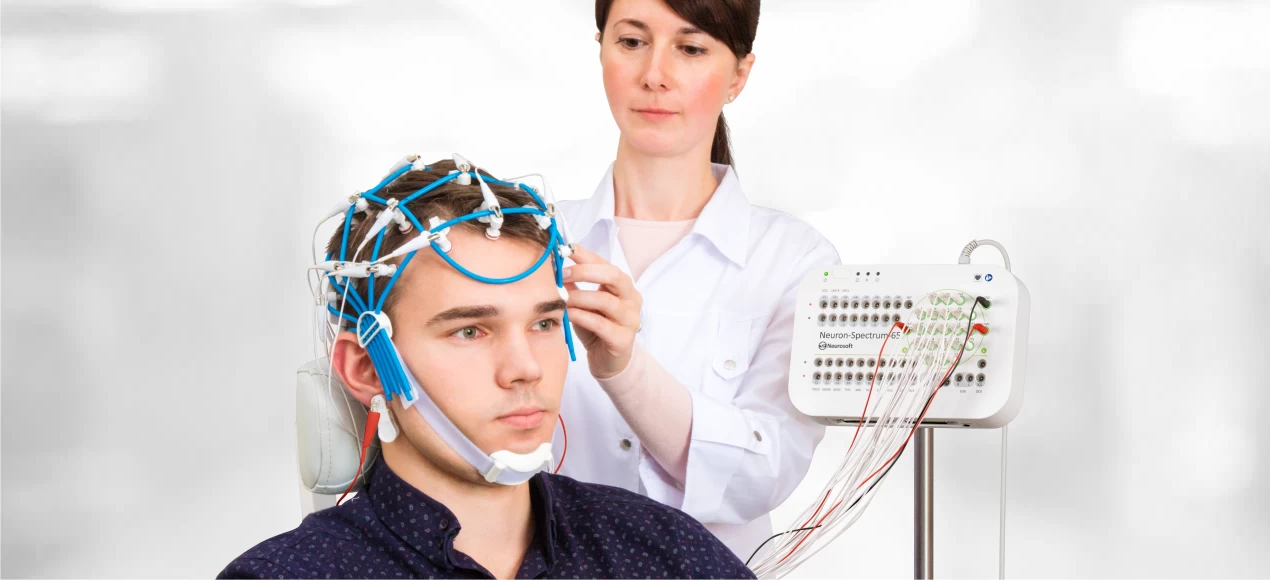
We recommend this expert class Neuron-Spectrum-65 system (can be used as Type I monitor according to AASM classification) for in-lab PSG.
EEG channels + differential channels to record any physiological signals
The device features 39 EEG channels, dedicated ECG, EMG and OEG channels, 8 additional differential channels and dedicated DC channels. The DC channels allow connecting any PSG sensors of third-party manufacturers as well as obtain data from PAP devices to determine therapeutic pressure in patients with OSA (obstructive sleep apnea).
Long-term video EEG monitoring
Thanks to the wide range of acquisition channels, Neuron-Spectrum-65 can be effectively used for long-term video EEG monitoring together with 32-channel electrode cap and for pre-operative monitoring together with other types of electrodes including invasive.
The monitoring may continue for up to several days or even weeks. The video can be recorded from 1, 2 or 3 cameras simultaneously and can be motion trigged. Mobile trolley-based configuration allows using it in ICU.
Polysomnography
Neuron-Spectrum-65/PSG configuration is a perfect solution for the most complex in-lab sleep studies performed in sleep centers: routine video PSG testing, MSLT (multiple sleep latency test), MWT (maintenance of wakefulness test), PSG with manual/automatic PAP titration, split-night PSG and others.
According to AASM, video monitoring synchronized with PSG data is recommended. HD cameras with IR illumination and remote control option ensure maximum convenience for the medical personnel when using the system. The video image helps accurately determine sleep and wake stages, and obtain a more complete clinical picture of patients with OSA, restless leg syndrome, parasomnia and other sleep-related disorders. Neuron-Spectrum.NET/PSG software module ensures manual, semi-automatic and automatic sleep staging and PSG event detection (apnea/hypopnea, snoring, desaturation, arrhythmia, limb movement, arousal, etc.). Automatic algorithms implemented in the software speed up PSG data processing saving precious time.
Portable patient unit for quick connection/disconnection
During in-lab PSG the sensors can be connected to the recorder through the portable patient unit. This allows a patient to disconnect quickly from the recorder (for example in case of bathroom needs) and then connect the sensors back to resume the recording.
Continuous impedance monitoring during acquisition
The color-coded impedance indication on the amplifier inputs and continuous impedance measurement during acquisition assist the specialist in signal quality monitoring and, if necessary, with timely correction of the electrode position thus ensuring superb recording quality. The operating mode can be switched at a push of a button on the front panel of the device whereas the LED indicator next to button will always show the current operating mode.


| • | EEG channel 32 + 9 additional channel (EMG, ECG, EOG, SpO2 .etc) |
| • | EMG & EP 4 Channel System |
| • | QEEG with 2 & 3D Mapping |
| • | QEEG Brain Mapping with Comparison database upto 82 Yrs |
| • | QEEG Result: Absolute Power, Relative Power, Coherence, Phase Lag, etc |
| • | Video EEG with synchronous Camera Full HD for LTM/ aEEG / PSG |
| • | Spike & Sharp Wave Detection |
| • | Epileptic Seizure Detection |
| • | Search Burst / Suppresion |
| • | aEEG / Amplitude Integrated EEG / CFM |
| • | The support of HL7 standard allows integrating all diagnostic neurosoft system |
| • | into the information system of a healthcare facility. |
| • | Impedance Measurement Button on the front panel. |
| • | Advance analysis tools |
| • | 3D Brain mapping and bar charts of EEG analysis results |
| • | Graph of spectral and coherence EEG analysis result |
| • | Automatic artefak detectio |
| • | Automatic spike detection and sharp waves |
| • | Creation and editing of EEG montage |
| • | Trend of EEG parameters |
| • | Automatic general EEG report |
| • | Compatible with loreta & MatLab |
| Amplifier | ||
| Number of Channel | : | 40+ channels |
| Voltage Range | : | 1 - 12000 μV |
| Ratio error Voltage Measurement | : | |
| in the range from 10 up to 50 μV | : | within ±25% |
| in the range from 50 up to 10000 μV | : | within ±7% |
| Sensitivity | : | 0.01 - 10.000.000 μV/mm |
| Ratio error of sensitivity | : | within ±5% |
| High pass filter | : | arbitary value can be set in 0.01 - 10 Hz range |
| Low pass filter | : | arbitary value can be set in 0.1 - 500 Hz range |
| Sweep Speed | : | arbitary value can be set in 1 - 10000 mm/s range |
| Ratio error sweep speed | : | within ±2% |
| Bandpass flatness in 0.5 - 60 Hz | : | from -10 up to +5% |
| Suppresion ratio of power frequency by notch filter | : | not less than 40 dB |
| CMRR | : | not less than 120 dB |
| Input noise level in 0.5 - 200 Hz range (rms) | : | not more than 1.4 μV |
| Input Impedance | : | not less than 400 MΩ |
| Patient leakage current | : | not more than 50 nA |
| Voltage measurement range | : | 0,2 - 100 mV |
| Flash Simulator | ||
| Flash Rate | : | 0 - 100 Hz |
| Manual, default or custom flash program | ||
| General Specifications and Parameters | ||
| Interface | : | USB |
| Supply voltage | ||
| electronic unit | : | 5 V DC |
| desktop PC based system | : | 220/230 V AC (50 Hz) |
| Electronic unit power consumption | : | not more than 2.8 VA |
| Safety | : | CF type |
| Synchronous Review EEG & Video | : | Include Camera Full HD |
| Operating Limitattions | ||
| Ambient temperature | : | 10 to 35 С |
| Relative humidity | : | 80 % at 25 C |
| Atmospheric pressure | : | 760±30 mmHg |
| Software | ||
| Export format avi, txt, rtf, xml, bmp, emf, EDF, pdf | ||
| Report automatically generated EEG report | ||
| EEG Software | ||
| Single / All channel editor | ||
| Single/ Multiple channel measurement | ||
| Magnifier Mode | ||
| Automatic detection of spikes and sharp waves | ||
| Automatic artifact detection | ||
| Bandpass Filter | : | Delta, Theta, Alpha, Beta, or custom rhytem |
| Brain Mapping / QEEG | : | 2D or 3D |
| Spectral Analysis | : | Spectrum graph, spectrum table, spectrum map |
| Coherence Analysis | ||
| EEG Trends | ||
| Wavelet analysis | ||
| EMG & EP Software | ||
| NCS | ||
| Motor and sensory condtion velocity, F-wave, Hreflex, motor and sensory conduction collision | ||
| EMG | ||
| Spontaneous activity, interference curve, MUP, Single Fiber & macro EMG | ||
| Neuromuscular Junction | ||
| MUNE | ||
| SSEP | ||
| VEP | ||
| AEP | ||
| Neuron-Spectrum-5 electronic unit | : | 1 |
| LED photic stimulator | : | 1 |
| Floor stand for LED photic stimulator | : | 1 |
| EEG Cup electrode with cable | : | 40 |
| Electrode Adhesive Paste | : | 3 |
| Abrasive Paste for Skin preparation | : | 3 |
| Electrode system Cap | : | 1 |
| User manual "Neuron-Spectrum.NET" soft copy | : | 1 |
| Equipment and Software for EMG: | ||
| Dedicated keyboard with cable and bluetooth adapter | : | 1 |
| Patient button (with USB connector) | : | 1 |
| USB hub with cable | : | 1 |
| Footswitch unit | : | 1 |
| Auditory stimulator (headphones) | : | 1 |
| Visual stimulator (LED goggles) | : | 1 |
| Stimulating bar electrode (adult) | : | 1 |
| Bar electrode (adult) | : | 1 |
| Bar electrode (pediatric) | : | 1 |
| Adjustable electro stimulating probe | : | 1 |
| Ground electrode with cable (pediatric) | : | 1 |
| Ground electrode with cable (adult) | : | 1 |
| Surface electrode | : | 2 |
| Ring electrode (wide) with cable | : | 1 |
| Crocodile Clip | : | 3 |
| Adapter for needle electrode connection | : | 1 |
| Disposable concentric EMG needle electrode | : | 25 |
| Disposable ECG electrode | : | 100 |
| Y-adapter | : | 2 |
| User manual "Neuro-MEP.NET" soft copy | : | 1 |
| License for EMG | : | 1 |
| License for EEG | : | 1 |
| License for QEEG Brain Mapping | ||
| Aksessoris Lokal | ||
| PC | : | 1 |
| Monitor | : | 1 |
| Printer | : | 1 |
| Trolley | : | 1 |
| UPS | : | 1 |
| Camera | : | 1 |
| Speaker | : | 1 |